
Testicular cancer
Testicular cancer is a malignant tumor that develops in the cells of one or, more rarely, both testicles. In more than 95% of cases, it originates from germ cells, i.e. those from which sperm cells are normally formed. Although it accounts for only about 1.6% of all malignant cancers in men, its importance increases dramatically in a certain age group. It is the most commonly diagnosed malignant neoplasm in young men, aged 20 to 44, accounting for as much as 25% of all oncological cases in this population.
Spis treści
Testicular Cancer – The Silent Enemy of Young Men and the Promise of Cure
The statistics for Polish are alarming. Every year, more than 1100 men are diagnosed, and over the last three decades, the incidence of this type of cancer has tripled. The peak of incidence falls between the ages of 20 and 39 – the time when men build their future, careers and start families.
It is in this context that the fundamental paradox of testicular cancer is revealed. On the one hand, it is a disease with an extremely dynamic and aggressive course, capable of metastasizing in just a few weeks. On the other hand, it is one of the cancers with the best prognosis. In the case of early detection, when the disease is limited only to the testicle, the cure rate reaches almost 100%. This duality – the enormous threat of delayed diagnosis and the extraordinary chance of full recovery with prompt intervention – is the key message of this guide. Knowledge about symptoms and regular self-examination are not only a recommendation, but the most important tool in the fight for life and health.
How to recognize testicular cancer? Key early symptoms that should not be ignored
The diagnosis of testicular cancer at an early stage is based on noticing subtle but characteristic changes. The key is to understand that the most typical symptom is often not associated with pain, which can be confusing and lull your vigilance.
Main and Most Common Symptom: Non-Painful Nodule
In about 90% of cases, the first alarm signal is the appearance of a hard, painless nodule in the testicle. This is the most important feature that should prompt immediate medical consultation. This tumor is palpable as a hard, rough lesion that is an integral part of the testicle – it cannot be separated or moved relative to its tissue. This distinguishes it from many benign lesions that are often located next to the testicle.
Changing the Size and Consistency of the Kernel
Another important symptom is noticeable enlargement, swelling or hardening of the entire testicle. The tumor testicle can become noticeably larger and heavier than the other. Sometimes, surprisingly, tumor development precedes earlier testicle shrinkage. Any change in its standard, springy consistency to a hard and uniform one is an alarm signal.
Feeling of Heaviness and Discomfort
Many men describe the first symptoms not as pain, but as an indefinable feeling of heaviness, expansion or “pulling” in the scrotum. This subtle discomfort, while easy to ignore in the daily rush, can be an early indicator of a developing tumor.
Testicular pain – a less typical, but important signal
The absence of pain is a psychological trap because it is commonly believed that a serious illness must hurt. In the case of testicular cancer, it is different – pain affects only one in four patients. His absence cannot be a reason to calm down. If the pain is already present, it may be dull and intermittent. Sudden and severe pain occurs rarely and may indicate complications within the tumor, such as bleeding into the tumor or ischemia.
Fluid buildup (reactive hydrocele)
Sometimes the body’s response to the presence of cancer is the accumulation of serous fluid in the testicular sheaths. This leads to the formation of the so-called reactive hydrocele, which manifests itself as enlargement and swelling of the scrotum. Hydrocele can mask the presence of a tumor, making it difficult to feel it, so any enlargement of the scrotum, even if it appears to be filled with fluid, requires ultrasound diagnostics.
Advanced Testicular Cancer: Metastatic Symptoms and Hormone Signals

Once the cancer cells have penetrated outside the nucleus, the disease enters an advanced stage. The symptoms are then no longer localized in the scrotum and begin to affect the entire body. Their appearance is a signal that the cancer has metastasized to other organs, which fundamentally changes the prognosis and intensity of the required treatment.
When cancer leaves the testicle – systemic symptoms
Testicular cancer most often spreads through the lymphatic and blood vessels in a predictable way. The location of metastases determines the type of symptoms:
- Retroperitoneal lymph node metastases: This is the first and most common direction of spreading. Enlarged lymph nodes deep in the abdominal cavity press on nerves and surrounding structures, causing persistent pain in the lower back (lumbar region) or abdominal pain. This symptom is often mistaken for sciatica or back problems.
- Lung metastases: Another common site of metastasis is the lungs. Respiratory symptoms include chronic cough, shortness of breath (feeling short of breath), chest pain, and in more advanced cases, even hemoptysis.
- Other rare metastases: In rarer cases, cancer cells can reach distant organs, giving specific symptoms. Bone metastases cause bone pain, while brain metastases can manifest themselves as headaches, dizziness or other neurological deficits.
Gynecomastia – An Unusual Signal from the Endocrine System
One of the most unusual and at the same time characteristic symptoms of testicular cancer is gynecomastia, i.e. enlargement and tenderness of the mammary glands in a man. This symptom occurs in about 10% of patients and is directly related to the hormonal activity of the tumor. Some types of testicular cancer, especially non-seminomas, produce a hormone called chorionic gonadotropin (beta-hCG) – the same hormone produced by women in early pregnancy. Its increased concentration in the man’s blood disturbs the hormonal balance, leading to the growth of the glandular tissue of the breast. The appearance of gynecomastia, especially unilateral or asymmetrical, in a young man should always be a signal to examine the testicles.
Testicular Self-Examination Step by Step: Your Most Important 5 Minutes a Month
Testicular self-examination is the most effective method of early detection of testicular cancer. It is a simple, painless and non-invasive procedure that takes just a few minutes and can save lives. Every man, starting from the age of 15, should do it regularly, once a month. The goal is not to obsessively search for cancer, but to build “somatic awareness” – to know your body well enough to immediately recognize any new, disturbing change, such as a painless enlargement of the testicle.
Preparation for the Study
- Best Time: Perform the test during or just after a warm bath or shower. The heat causes the scrotal muscles to relax, making the testicles hang freely and are easier to examine.
- The right conditions: Stand in a well-lit place, preferably in front of a mirror. This will allow for an accurate visual assessment of the scrotum, which is important in the context of detecting a testicular tumour.
Self-Examination Instruction (Detailed Steps)
- Visual inspection: Stand in front of a mirror and carefully examine the skin of the scrotum. Pay attention to any changes in its appearance – unusual redness, swelling, skin changes or deformities.
- “Weighing” in Hands: Gently grasp the scrotum with both hands and assess the weight of the testicles. Pay attention to whether one of them does not seem unnaturally heavier than during the previous examination.
- Palpation: Examine each kernel separately. Use your index and middle fingers from the bottom and your thumb from the top. Gently but firmly, roll the testicle between your fingers, examining its entire surface. Look for any hard bumps, lumps or bumps.
- What do you sense? A healthy kernel should be smooth, oval-shaped, and have a uniform, springy consistency, similar to a hard-boiled egg without a shell. On the back and top of each testicle, you will feel a soft, oblong structure – this is the epididymis where sperm are stored. It is a completely normal part of the anatomy and should not be confused with a tumor, however, cryptorchidism can increase the risk of developing testicular cancer.
What should you be concerned about?
- Feeling a hard, non-movable lump or lump on the surface or inside the testicle.
- Any change in size, shape, or consistency (hardness) of the testicle compared to previous studies.
- The appearance of pain or discomfort when touching that has not occurred before.
- Remember: A slight difference in the size of the testicles (one is usually slightly larger and lower-hung) is completely normal.
Regularity is the key
For self-examination to be effective, it must become a habit. Do it once a month, preferably on the same day (e.g. to detect a testicular tumor early). on the first Saturday of the month, when tests for the occurrence of testicular cancer take place. This will help you learn what your testicles look like and how they feel, and any change will be easy to spot. If you detect anything alarming, don’t panic, but don’t delay – make an appointment with a doctor immediately.
A lump in the scrotum – is it really cancer? Other possible causes
Detecting a lump or other abnormality in the scrotum is always a cause for concern, but it’s important to remember that not every change is cancerous. There are a number of other, much more common conditions that can cause similar symptoms. Still, the key rule is that any palpable tumor in the scrotum requires urgent consultation with a doctor (general practitioner or urologist) in order to make the correct diagnosis.
The most important feature that can suggest cancer on physical examination is its inseparable connection with testicular tissue. A cancerous tumor is part of the testicle itself. Many benign lesions, such as cysts or inflammation, affect structures adjacent to the testicle (e.g. epididymis) and can be distinguished from the testicle itself on palpation.
Testicular cancer vs. other scrotal diseases – key differences
| Feature | Testicular cancer | Epididymitis/orchitis | Hydrocele testicular | Varicoceles |
| Pain | Usually painless (pain in about 25% of patients) | Usually severe, sharp pain, increasing rapidly | Usually painless, feeling heavier | Usually painless, sometimes a feeling of pulling |
| Consistency | Hard, solid tumor, uneven surface | Swelling and tenderness, testicle and epididymis painful to touch | Soft, resilient bump A fluid-filled tumor in the testicle can be a sign of cancer, so it’s important to see a doctor. | Soft, winding structures, described as a “bag of worms” |
| Location | The tumor is an integral part of the testicle, it cannot be separated from the context of symptoms and treatment of testicular cancer. | Inflammation involves the epididymis and/or testicle | Fluid accumulates around the entire testicle | Dilated veins located above the testicle |
| Accompanying symptoms | Possible gynecomastia, symptoms of metastasis (back pain, cough) | Fever, redness of the scrotum skin, symptoms of urinary tract infection | Enlargement of the scrotum may be a symptom of painless enlargement of the testicle, which requires further diagnosis., possible transillumination (passage of light) | The change is palpable mainly in the standing position, but may disappear in the lying position |
Diagnostic pathway: how does the doctor make the diagnosis?
The diagnostic process in the case of suspected testicular cancer is fast, precise and based on standard procedures. Its purpose is not only to confirm or exclude cancer, but also to determine its stage at the same time, which is crucial for planning further treatment.
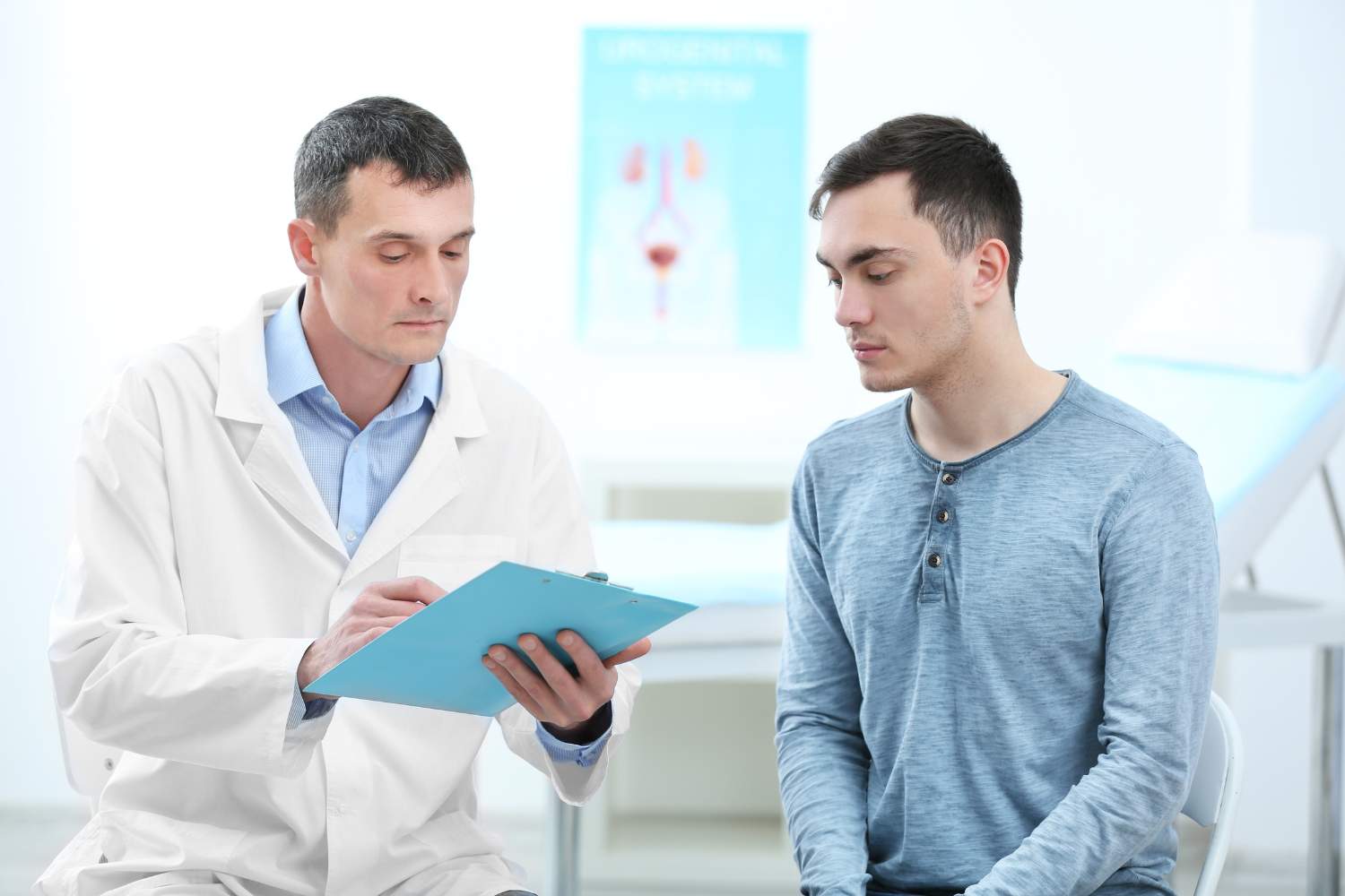
First Step: Visit to the Doctor
It all starts with a visit to the general practitioner or directly to the urologist. The doctor will conduct a detailed medical history and physical examination (palpation), during which he will assess the size, shape, consistency and tenderness of both testicles and scrotum. If abnormalities are found, the patient will be urgently referred for further tests.
Imaging test: ultrasound of the scrotum
Ultrasound of the scrotum is the gold standard and a key test in the diagnosis of testicular tumors. It is a completely painless, non-invasive and quick procedure. With the use of sound waves, it allows to visualize the structure of the testicle with almost 100% sensitivity and distinguish a solid tumor (suspected of cancer) from fluid lesions, such as a cyst or hydrocele of the testicle.
Blood tests: tumor markers
In parallel with the ultrasound, the doctor will order blood tests to determine the concentration of three key tumor markers. Their level is extremely important in diagnosis, assessment of prognosis and monitoring of treatment effectiveness.
- AFP (Alpha-Fetoprotein): It is a protein whose level is very low in healthy adult men. Its concentration is elevated in some types of non-seminomas. Importantly, in the case of pure seminomas, AFP levels always remain normal, making it an important differential marker.
- Beta-hCG (Chorionic Gonadotropin): This hormone can be produced by both seminomas and non-seminomas, although it is more common and in higher concentrations in the latter. Normal levels of beta-hCG in men should not exceed 2-5 mIU/ml, depending on the laboratory.
- LDH (Lactate Dehydrogenase): It is an enzyme present in many cells of the body, which is why it is a non-specific marker. However, its significantly elevated level in the case of a confirmed testicular tumor may indicate a large tumor mass and the advancement of the disease.
Tumor Markers in Testicular Cancer – Interpretation and Meaning
| Marker | Indicative Norm in Men | The importance of elevated levels | In what types of cancer does it occur? The most common cancer in young men is testicular cancer. |
| AFP | <7 ng/ml | It indicates the presence of a non-seminoma component. | Only non-seminomas (e.g. germ cell carcinoma, yolk bladder tumor). |
| Beta-hCG | <2−5 mIU/ml | It indicates the presence of active cancer cells and correlates with the tumor mass. | Non-seminomas (40-60% of cases), seminomas (15-20% of advanced cases). |
| LDH | Laboratory-dependent, tumor marker analysis may indicate the presence of testicular cancer. | A non-specific indicator of high tumor weight, rapid growth, and disease advancement. | Seminomas (about 70% of cases), non-seminomas (20-70% of cases). |
Final confirmation – histopathological examination
It should be emphasized that the final and 100% diagnosis of the type of cancer and its histological features is possible only after surgical removal of the testicle (a procedure called orchiectomy) and examination of the tumor under the microscope by a pathomorphologist. Due to the high risk of cancer cell dissemination, if testicular cancer is suspected, a needle biopsy of the tumor is usually not performed before its removal.
Types and Stages of Testicular Cancer
Once the diagnosis is confirmed, it is crucial to determine two parameters for planning the treatment of testicular cancer: the histological type of the cancer and its clinical stage, which often requires histopathological examination of the testicle.
Two main types of germ cell tumors
Testicular germ cell tumors, accounting for more than 95% of all cases, are divided into two main groups, which differ in biology, course and response to treatment. The decision whether a given tumor is seminoma or non-seminoma is one of the most important crossroads in the patient’s therapeutic path.
- Seminomas: They account for 25% to 50% of cases. They usually develop more slowly and have less tendency to metastasize early. They occur in slightly older men, most often aged 25-45. Their key feature is very high sensitivity to radiotherapy, which is one of the main options for adjuvant treatment.
- Non-seminomas: They account for 50% to 75% of cases and predominate in younger men, aged 15-30 years. This is a more diverse group, which includes, m.in others, reproductive cells, associated with the occurrence of testicular cancer. germ cell carcinoma, teratoma, chorionoma or yolk sac tumor. Non-seminomas grow faster, are more aggressive and metastasize more often compared to the most common cancers in young men. Unlike seminomas, they are resistant to radiation therapy, but respond very well to chemical treatment (chemotherapy).
Staging
The severity determines how much the disease has spread in the body. A simple, three-stage classification is used for this:
- Grade I: The tumor is limited to the testicle only. There is no evidence of metastases in imaging tests (e.g. CT scan) or in the blood (tumor markers return to normal after surgery), but it is important to examine the testicles yourself and see a doctor in case of worrying symptoms.
- Grade II: The disease has spread beyond the testicle to lymph nodes located in the retroperitoneal space (deep in the abdominal cavity). There are no metastases in distant organs.
- Grade III: The presence of distant metastases is found – in lymph nodes outside the abdominal cavity (e.g. in the mediastinum, above the clavicle) or in other organs, such as the lungs, liver, bones or brain.
Testicular Cancer Treatment: Effective and Proven Methods
Treatment of testicular cancer is one of the greatest successes of modern oncology. Thanks to the combination of surgery, chemotherapy and radiotherapy, it is a disease with a very high cure rate, even in advanced stages. Therapy is always carried out by a multidisciplinary team of specialists (urologist, clinical oncologist, radiotherapist) in specialized centers.
Basis of treatment: inguinal orchiectomy
The first and absolutely necessary step in the treatment of any testicular cancer is its surgical removal. This procedure, called radical inguinal orchiectomy, involves the removal of the entire testicle along with the epididymis and spermatic cord. What is very important, the operation is performed from a small incision in the groin, not through an incision on the scrotum. This approach minimizes the risk of cancer cell dissemination and is the standard oncological management for tumors in the testicle. The removed testicle is then sent for histopathological examination, which finally determines the type of cancer.
Adjuvant (adjuvant) treatment
After the surgery, depending on the histological type of the tumor and the stage of the disease, adjuvant treatment is implemented. Its purpose is to destroy possible micrometastases and reduce the risk of recurrence.
- Chemotherapy: It is the basis of systemic treatment, especially in the case of non-seminomas and in the advanced stage of testicular cancer and in both types of cancer. Modern chemotherapy for testicular cancer is based on regimens containing cisplatin (e.g. BEP regimen: bleomycin, etoposide, cisplatin), which has revolutionized the treatment of this disease and drastically improved the prognosis.
- Radiotherapy: It is the method of choice in the adjuvant treatment of stage I and II seminomas. It involves precise irradiation of the retroperitoneal lymph nodes with ionizing rays to destroy potential metastases. Seminomas are extremely sensitive to this form of therapy.
- Retroperitoneal lymphadenectomy (RPLND): It is a major surgical procedure involving the removal of lymph nodes from the retroperitoneal space. It is mainly used in the treatment of non-seminomas, as an alternative to chemotherapy in some stage I cases, or to remove residual tumor masses after chemotherapy.
Prognosis – very good forecasts
Testicular cancer, despite its aggressive nature, is characterized by an excellent prognosis. The effectiveness of treatment is very high, provided that it is undertaken in a timely manner.
- In stage I, in the context of testicular cancer , when the cancer is confined to the testicle, the 5-year survival rate ranges from 97% to almost 100%.
- Even in the case of metastatic disease (stage II and III), thanks to modern chemotherapy, cure rates are impressive and reach 70-80%.
These data clearly show that the diagnosis of “testicular cancer” is not a sentence, but the beginning of the road to full recovery.
Life After Testicular Cancer: A Return to Full Health and Masculinity
Oncological treatment is not only a fight against cancer, but also a process of adaptation to the new reality. Modern medicine approaches the patient in a holistic way, offering support in fertility, sexual health, mental health and body aesthetics. These aspects are not add-ons, but a standard element of comprehensive care that every patient should actively ask their treatment team about.
Fertility – A Key Issue to Address Before Treatment
- Oncofertility and Sperm Banking: Chemotherapy and radiation, while life-saving, can irreversibly damage sperm-producing cells in a healthy testicle, leading to permanent infertility. Therefore, the absolute priority for every man before starting adjuvant treatment is fertility preservation. The simplest and most effective method is to deposit sperm in a sperm bank (cryopreservation). It is a quick procedure, lasting only 1-2 days, which does not delay oncological treatment. In Poland, from June 1, 2024, this procedure is reimbursed for oncology patients.
- Important information: It should be emphasized that the removal of one testicle (orchiectomy) alone does not cause infertility. The second, healthy testicle is able to fully take over the function of sperm and hormone production, allowing the child to conceive naturally.
Sexual Performance and Hormones
Fears of losing masculinity are natural, but in most cases unfounded. Removal of one testicle and standard oncological treatment usually do not adversely affect the ability to achieve an erection or libido. Sometimes, especially after the removal of both testicles or as a result of treatment, testosterone levels can be reduced. This manifests itself in fatigue, low mood and reduced sex drive. It is a condition that can be easily diagnosed with blood tests and effectively treated with hormone replacement therapy (administration of testosterone in the form of injections or gel).
Mental Health and Psycho-Oncological Support
A cancer diagnosis is a huge psychological burden. Anxiety, uncertainty, sadness, and even depression are natural reactions to such a difficult situation. It is crucial not to be left alone with these emotions, especially in the face of the risk of developing testicular cancer. The support of a psycho-oncologist, i.e. a psychologist specializing in working with oncological patients, is an invaluable part of the treatment process. It helps to tame anxiety, cope with stress, communicate with loved ones and the medical team, and find oneself in the new reality. Asking for psychological help is not a sign of weakness, but of maturity and responsibility for one’s full health.
Reconstruction and Appearance – Implantation of a Testicular Prosthesis
For many men, testicle loss is associated with concerns about appearance and self-attractiveness. Modern surgery offers a simple and effective solution to this problem – the placement of a testicular implant (prosthesis). It is a silicone prosthesis with a shape and consistency similar to a natural testicle, which is placed in the scrotum. This procedure allows for the restoration of symmetry and natural appearance, which significantly improves the patient’s psychological comfort and self-esteem. Implantation can be performed at the same time as the surgery to remove the testicle or at a later date, after the entire oncological treatment is completed.
Post-Treatment Check-Ups: Long-Term Health Care
The end of treatment is the beginning of a new stage – regular observation. Check-ups are a key part of post-cancer care. Their goal is to detect a possible recurrence early, monitor the condition of the second testicle and possible long-term side effects of therapy, such as cardiac or metabolic problems.
Schedule of Visits and Scope of Examinations
The period of active oncological observation usually lasts from 5 to 10 years, which is important in the context of testicular cancer prevention. The frequency of examinations is highest in the first two years after treatment, when the risk of recurrence is highest, and then gradually decreases. Modern control schemes seek a balance between oncological vigilance and minimizing the burden on the patient, e.g. by limiting the doses of ionizing radiation from frequent computed tomography scans in favor of other tests, such as magnetic resonance imaging (MRI).
The standard control scheme includes:
- A doctor’s visit should be done regularly, especially in the context of cancer prevention. Interview and physical examination.
- Determination of tumor markers: Regular blood tests for AFP, beta-hCG and LDH levels.
- Imaging tests: Computed tomography (CT) scan of the abdomen and chest, chest X-ray or MRI, according to an individual schedule set by the doctor.
- Self-examination: The patient should continue monthly self-examination of the other, healthy testicle.
The table below presents a simplified, exemplary control scheme for patients after treatment of stage I testicular cancer. It is important to remember that this is only a general outline and the exact treatment plan is always determined individually by the treatment team, taking into account the risk factors.
Simplified Follow-Up Schedule After Stage I Testicular Cancer Treatment
| Post-treatment period | Doctor’s visit + Tumor markers | Imaging tests (CT/MRI/X-RAY) |
| Year 1-2 | Every 2-4 months | According to the schedule, e.g. 2-4 times per period |
| Year 3-5 | Every 6-12 months | On schedule, e.g. 1 time a year |
| After 5 years | Usually 1 time a year | Depending on the indications and policy of the center |
Your health, your responsibility, your win
Testicular cancer is a disease full of contrasts: it is aggressive, but also highly curable; Its main symptom is insidiously painless, but also easy to detect on your own. The key to overcoming this cancer is time. Each day of delay in diagnosis reduces the chances of simple and effective treatment, while a quick response to a disturbing symptom almost guarantees full recovery.
Your most important ally in this fight is your own hand and the regularity of your monthly self-examination. It’s a simple activity that puts you in control of your own health. Remember that any disturbing change, lump, enlargement or hardening of the testicle requires immediate consultation with a doctor. There is no place for shame or procrastination here.
Modern medicine offers extremely effective methods of treating testicular cancer and comprehensive support at every stage of the disease – from fertility protection, through psychological assistance, to aesthetic reconstruction. Diagnosis is not the end of the world, but the beginning of a road on which you have science, experienced specialists and growing social awareness on your side. Your health is in your hands. Be brave, be vigilant, win life.